Viral infections pose significant threats to plant health, impacting agriculture and medicinal plant production. This study focuses on diagnosing viral infections in Ipomoea cairica (L.) Sweet using advanced techniques such as Transmission Electron Microscopy (TEM) and Microtomy. Phenotypic symptoms, anatomical changes, and the confirmation of Gemini virus through TEM are investigated. Transmission studies reveal multiple vectors, including Bemisia tabaci, highlighting the importance of understanding viral spread mechanisms. Host range studies demonstrate widespread susceptibility among various plant families. The research emphasizes early diagnosis and management to mitigate economic losses and preserve medicinal metabolites. Future efforts will employ PCR and DNA sequencing for precise viral strain identification and targeted management strategies. The findings underscore the importance of early diagnosis and management of viral infections to safeguard plant health and preserve essential metabolites. Future research aims to utilize PCR and DNA sequencing for precise identification and targeted management of viral strains, enabling effective disease control strategies for Ipomoea cairica (L.) Sweet and similar plant species. This study underscores the necessity for proactive measures to protect plant species like Ipomoea cairica (L.) Sweet from viral infections, ensuring sustainable agriculture and medicinal plant production.
| Published in | International Journal of Microbiology and Biotechnology (Volume 9, Issue 2) |
| DOI | 10.11648/j.ijmb.20240902.11 |
| Page(s) | 30-36 |
| Creative Commons |
This is an Open Access article, distributed under the terms of the Creative Commons Attribution 4.0 International License (http://creativecommons.org/licenses/by/4.0/), which permits unrestricted use, distribution and reproduction in any medium or format, provided the original work is properly cited. |
| Copyright |
Copyright © The Author(s), 2024. Published by Science Publishing Group |
Morphological, Histopathological Studies, Viral Infections, Electron Microscopy
2.1. Sample Collection
2.2. Phenotypic Symptoms of the Disease
2.3. Histological Sectioning Using Microtome
2.4. Transmission Electron Microscopy of Infected Sample
2.5. Transmission Studies of Viral Isolate
2.6. Host Range Studies of Viral Isolate
3.1. Histological and Anatomical Effect
3.2. Histology of Healthy Leaves
3.3. Histology of Infected Leaves
3.4. Transmission Electron Microscopy (TEM)
3.5. Transmission Studies
Method of Transmission | No. of plants inoculated | No. of plant infected | Incubation Period (Days) | Transmission rate |
|---|---|---|---|---|
Bain’s Method | 10 | 5 | 20 | 50% |
Matz’s Method | 10 | 4 | 20 | 40% |
Combined method | 10 | 6 | 20 | 60% |
Aphids | Plant infected/ Plant exposed | % Transmission |
|---|---|---|
Aphis craccivora | 2/20 | 10% |
Aphis gossypii | 6/20 | 30% |
Aphis nerii | 2/20 | 10% |
Bemisia tabaci | 18/20 | 90% |
Myzus persicae | 2/20 | 10% |
3.6. Host Ranges Studies of Viral Isolate
S.No | Host Species | Family | Response-Positive (+) or Negative (-) | Symptoms |
|---|---|---|---|---|
1. | Abelmoschus esculentus (Bhindi) | Malvaceae | + | Necrotic Spots |
2. | Ageratum Spp. (Whiteweed) | Asteraceae | + | Leaf deformation |
3. | Benincasa hipsida (Wax gourd) | Cucurbitaceae | + | Leaf deformation |
4. | Cucumis melo (Melon) | Cucurbitaceae | + | Vein clearing |
5. | Cucumis sativa (Cucumber) | Cucurbitaceae | + | Vein clearing |
6. | Capsicum annum (Chillies) | Solanaceae | + | Leaf deformation |
7. | Carica papaya (Papaya) | Cairicaceae | + | Chlorotic spots and leaf deformation |
8. | Citrullus lanatus (Watermelon) | Cucurbitaceae | + | Mild Mosaic |
9. | Cucumis pepo (Pumpkin) | Cucurbitaceae | + | Vein clearing |
10. | Cyamopsis tetragonoloba (Guar) | Fabaceae | + | Blistering of leaf lamina |
11. | Catharanthus roseus (Sadabahar) | Apocynaceae | + | Mild Mosaic, colour breaking petals |
12. | Chenopodium album (Baconweed) | Amaranthaceae | + | Systemic lethal necrosis |
13. | Coccinia grandis (Ivy gourd) | Cucurbitaceae | + | Leaf deformation |
14. | Cucucurbita maxima (Winter squash) | Cucurbitaceae | + | Vein clearing |
15. | Calatropis procera (Madaar) | Apocynaceae | + | Systemic lethal necrosis |
16. | Convolvulus arevensis (bindweed) | Convolvulaceae | + | Mild mosaic |
17. | Cucurbita vulgaris (Squash) | Cucurbitaceae | + | Vein clearing |
18. | Chrysanthemum indicum (Chrysanthemum) | Asteraceae | + | Mild mosaic |
19. | Crossandra infundibulum (Firecracker) | Acanthaceae | + | Blistering of leaf lamina |
20. | Daucus carota (Wild carrot) | Apiaceae | + | Necrotic local lesion |
21. | Glycine max (Soyabean) | Fabaceae | + | Leaf deformation |
22. | Gossypium hirsutum (Cotton) | Malvaceae | + | Chlorotic Spots |
23. | Hibiscus cannabinus (Kenaf) | Malvaceae | + | Blistering of leaf lamina, Mild mosaic |
24. | Jatropha spp. (Physic nut) | Euphorbiaceae | + | Vein clearing and bending |
25. | Jasminum multiflora (Star jasmine) | Oleaceae | + | Necrosis |
26. | Lagenaria siceraria (Bottle gourd) | Cucurbitaceae | + | Vein clearing |
27. | Luffa cyclindrica (Sponge gourd) | Cucurbitaceae | + | Vein clearing |
28. | Lycopersicon esculentum (Tomato) | Solanaceae | + | Mild mosaic |
28. | Momordica charantia (Bitter gourd) | Cucurbitaceae | + | Vein clearing |
29. | Papaver somnifera (Poppy) | Papaveraceae | + | Blistering of leaf lamina |
30. | Solanum nigrum (Black night shade) | Solanaceae | + | Mild mosaic |
31. | Solanum melongena (Eggplant) | Solanaceae | + | Mild mosaic |
32. | Solanum tuberosum (Potato) | Solanaceae | + | Mild mosaic |
33. | Sauropus androgynus (Star gooseberry) | Phyllanthaceae | + | Vein clearing |
34. | Trichosanthes cucumerina (Snakegourd) | Cucurbitaceae | + | Leaf deformation |
35. | Triticum aestivum (Wheat) | Poaceae | - | No Symptoms |
36. | Tagetus erectus (Marigold) | Asteraceae | - | No Symptoms |
37. | Cynodon dactylon (Scutch grass) | Poaceae | - | No symptoms |
38. | Lantana indica (West Indian Lantana) | Verbenaceae | - | No Symptoms |
39. | Pennisetum pedicillatum (Grassweed) | Poaceae | - | No symptoms |
40. | Zea mays (Maize) | Poaceae | - | No Symptoms |
| [1] | Mettenleiter T. C. (2017). “The first virus hunters,” in Advances in Virus Research, Vol. 99, eds M. Beer and D. Höper (Burlington: Academic Press): 1–16. |
| [2] | Koonin E. V., Dolja V. V., and Krupovic M. (2015). Origins and evolution of viruses of eukaryotes: the ultimate modularity. Virology; 479–480: 2–25. |
| [3] | Gentile M., and Gelderblom H. R. (2014). Electron microscopy in rapid viral diagnosis: an update. New Microbiol, 37: 403–422. |
| [4] | King A. M. Q., Lefkowitz E., Adams M. J., and Carstens E. B. (2012). Ninth Report of the International Committee on Taxonomy of Viruses. Elsevier. |
| [5] | Austin D. F. and Huaman Z. (1996). A synopsis of Ipomoea (Convolvulaceae) in the Americas. Taxon, 45: 5. |
| [6] | Syller (2014). Biological & Molecular events associated with simultaneous transmission of plant viruses by invertebrate and fungal vectors. Mol. Plant Pathol, 15(4): 417-426. |
| [7] | Valverde R. A., Clark C. A., Valkonen J. P. T. (2007). Viruses and Virus Disease complexes of Sweetpotato. Plant Viruses, 1(1): 116-126. |
| [8] | Sivparsad B. J., Gubba A. (2013). Identification and Distribution of viruses infecting Sweet Potato (Ipomoea batatus) in KwaZulu- Natal, South Africa. South African Journal of Plant and Soil, 30(3): 179-190. |
| [9] | Mohammed F., Arishiya T. F., Mohamed S. (2012). Microtomes & Microtome knives- A Review & proposed classification. Annals Dent. Univ. Malaya; 19(2): 43-50. |
| [10] | Adekulne OK., Kulshrestha S., Prasad R., Hallan V., Raikhy G., Verma N., Ram R., Kumar S., and Zaidi AA. (2005). Plant-parasitic and vector nematodes associated with Asiatic and oriental hybrid lilies. Bioresources Technology. |
| [11] | Sass J. E. (1951) Botanical microtechnique Iowa state university Press, Ames. 228pp. |
| [12] | Esau K. (1938). Some anatomical aspects of plant virus diseases problem. I. Bot. Rev. 4: 548-579. |
| [13] | Martelli G. P., and Russo M. (1977). Plant virus inclusion bodies. Adv. Virus Res. 12: 175-266. |
| [14] | Khurana SMP. (1968). A study of virus disease of papaya (Cairica Papaya L.) in Gorakhpur. Ph.D. thesis. Univ of Gorakhpur. |
| [15] | Purcifull D. E., Edwardson J. R. and Christle S. R. (1968). Aggregated Filaments in extracts of Lupinus infected with watermelon mosaic virus. Virology. 35: 478-482. |
| [16] | Nault L. R. (1997). Arthropod transmission of plant viruses: A new synthesis. Ann. Entomol. Soc. Am. 90: 521-541. |
| [17] | Ng J. C. K., and Falk BW. (2006). Virus vector interactions mediating non-persistent and semi-persistent transmission of plant viruses. Annv. Rev Phytopathil. 44: 183-212. |
| [18] | Levy, D., Marco, S., 1982. The quality of seed tuber in the Golien Heights for spring season in Israel. Potato Res 25: 107-1597. London, Vol 1, p 384. |
APA Style
Rauniyar, N., Srivastava, D. (2024). Electron Microscopic and Histopathological Studies of the First Viral Infection Reported in Ipomoea Cairica (L.) Sweet. International Journal of Microbiology and Biotechnology, 9(2), 30-36. https://doi.org/10.11648/j.ijmb.20240902.11
ACS Style
Rauniyar, N.; Srivastava, D. Electron Microscopic and Histopathological Studies of the First Viral Infection Reported in Ipomoea Cairica (L.) Sweet. Int. J. Microbiol. Biotechnol. 2024, 9(2), 30-36. doi: 10.11648/j.ijmb.20240902.11
AMA Style
Rauniyar N, Srivastava D. Electron Microscopic and Histopathological Studies of the First Viral Infection Reported in Ipomoea Cairica (L.) Sweet. Int J Microbiol Biotechnol. 2024;9(2):30-36. doi: 10.11648/j.ijmb.20240902.11
@article{10.11648/j.ijmb.20240902.11,
  author = {Neha Rauniyar and Deepa Srivastava},
  title = {Electron Microscopic and Histopathological Studies of the First Viral Infection Reported in Ipomoea Cairica (L.) Sweet
},
  journal = {International Journal of Microbiology and Biotechnology},
  volume = {9},
  number = {2},
  pages = {30-36},
  doi = {10.11648/j.ijmb.20240902.11},
  url = {https://doi.org/10.11648/j.ijmb.20240902.11},
  eprint = {https://article.sciencepublishinggroup.com/pdf/10.11648.j.ijmb.20240902.11},
  abstract = {Viral infections pose significant threats to plant health, impacting agriculture and medicinal plant production. This study focuses on diagnosing viral infections in Ipomoea cairica (L.) Sweet using advanced techniques such as Transmission Electron Microscopy (TEM) and Microtomy. Phenotypic symptoms, anatomical changes, and the confirmation of Gemini virus through TEM are investigated. Transmission studies reveal multiple vectors, including Bemisia tabaci, highlighting the importance of understanding viral spread mechanisms. Host range studies demonstrate widespread susceptibility among various plant families. The research emphasizes early diagnosis and management to mitigate economic losses and preserve medicinal metabolites. Future efforts will employ PCR and DNA sequencing for precise viral strain identification and targeted management strategies. The findings underscore the importance of early diagnosis and management of viral infections to safeguard plant health and preserve essential metabolites. Future research aims to utilize PCR and DNA sequencing for precise identification and targeted management of viral strains, enabling effective disease control strategies for Ipomoea cairica (L.) Sweet and similar plant species. This study underscores the necessity for proactive measures to protect plant species like Ipomoea cairica (L.) Sweet from viral infections, ensuring sustainable agriculture and medicinal plant production.
},
 year = {2024}
}
TY  - JOUR
T1  - Electron Microscopic and Histopathological Studies of the First Viral Infection Reported in Ipomoea Cairica (L.) Sweet

AU  - Neha Rauniyar
AU  - Deepa Srivastava
Y1  - 2024/04/02
PY  - 2024
N1  - https://doi.org/10.11648/j.ijmb.20240902.11
DO  - 10.11648/j.ijmb.20240902.11
T2  - International Journal of Microbiology and Biotechnology
JF  - International Journal of Microbiology and Biotechnology
JO  - International Journal of Microbiology and Biotechnology
SP  - 30
EP  - 36
PB  - Science Publishing Group
SN  - 2578-9686
UR  - https://doi.org/10.11648/j.ijmb.20240902.11
AB  - Viral infections pose significant threats to plant health, impacting agriculture and medicinal plant production. This study focuses on diagnosing viral infections in Ipomoea cairica (L.) Sweet using advanced techniques such as Transmission Electron Microscopy (TEM) and Microtomy. Phenotypic symptoms, anatomical changes, and the confirmation of Gemini virus through TEM are investigated. Transmission studies reveal multiple vectors, including Bemisia tabaci, highlighting the importance of understanding viral spread mechanisms. Host range studies demonstrate widespread susceptibility among various plant families. The research emphasizes early diagnosis and management to mitigate economic losses and preserve medicinal metabolites. Future efforts will employ PCR and DNA sequencing for precise viral strain identification and targeted management strategies. The findings underscore the importance of early diagnosis and management of viral infections to safeguard plant health and preserve essential metabolites. Future research aims to utilize PCR and DNA sequencing for precise identification and targeted management of viral strains, enabling effective disease control strategies for Ipomoea cairica (L.) Sweet and similar plant species. This study underscores the necessity for proactive measures to protect plant species like Ipomoea cairica (L.) Sweet from viral infections, ensuring sustainable agriculture and medicinal plant production.

VL  - 9
IS  - 2
ER  -
Department of Botany, Deen Dayal Upadhyay Gorakhpur University, Gorakhpur, India
Department of Botany, Deen Dayal Upadhyay Gorakhpur University, Gorakhpur, India
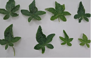
Figure 1. Demonstration of Infection based on Phenotypic symptoms of Ipomoea cairica (L.) Sweet.
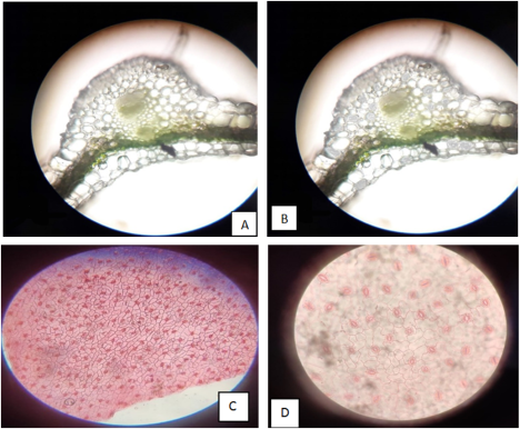
Figure 2. Histological demonstration of Ipomoea cairica (L.) Sweet. (A) Section of infected Leaf. (B) Section of Infected leaf. (C) Numerous stomata in infected leaf. (D) Reduced stomata in healthy leaf.
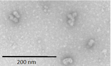
Figure 3. Particles of Gemini virus stained in Uranyl acetate showing typical twinned quasi-isometric subunit. The bar represents 200nm.