Research Article | | Peer-Reviewed

Assessment of Saliva and Urine Performance for Antimalarial Drug Resistance Molecular Markers Study

Received: 7 July 2024     Accepted: 2 August 2024     Published: 20 August 2024
Views:       Downloads:
Abstract

Background: The malaria diagnostic tools developed to date require blood to be taken. However, certain groups in the population are reluctant to take blood samples because of their cultural habits (blood taboo), or because of the fear associated with the trauma of the injection, especially when the sample is taken repeatedly. Saliva and urine, which are not very invasive to collect, have not been widely used for malaria diagnosis. The aim of this study is to assess the performance of saliva and urine in detecting molecular markers of Plasmodium falciparum resistance to antimalarial drugs. Methodology: Blood, urine and saliva samples were collected in three different localities from 94 patients over 2 years of age with microscopically confirmed Plasmodium falciparum uncomplicated malaria. P. falciparum genomic DNA (Deoxyribonucleic acid) was then extracted and amplified using primers specific for the Pfcrt (Plasmodium falciparum Chloroquine Resistance Transporter), Pfdhfr (Plasmodium falciparum dihydrofolate reductase) and PfK13 propeller (Plasmodium falciparum Kelch13 propeller) genes. The amplification products were processed by electrophoresis and analyzed against blood, saliva and urine samples. A multivariate statistical analysis in R programming environment was performed aiming to assess the performance of blood, saliva and urine samples in detecting molecular markers of P. falciparum resistance. Results: Agarose gel electrophoresis of the amplification products of each gene detected the Pfcrt genes at 80.85% (76/94), Pfdhfr at 95.74% (90/94) and PfK13 Propeller at 98.93% (93/94) in blood. In saliva, gene detection levels were 50% (47/94), 69.14% (65/94) and 4.26% (4/94) respectively for the K13 propeller, Pfdhfr and Pfcrt genes. Unlike the Pfcrt gene, which was not detected, 45.74% (43/94) and 38.30% (36/94) of PfK13 Propeller and pfdhfr genes respectively were detected in urine. Taking blood as the reference biological sample, statistical analysis showed that unlike urine, saliva exhibited a detection performance for molecular markers of antimalarial drug resistance (pfcrt, pfdhfr, pfK13 propeller) close to that of blood (p < 0.05). The performance of saliva and urine was also assessed on the basis of the detection of the molecular markers pfdhfr, pfcrt and pfK13 using ROC (receiver operational characteristic) analysis. The data revealed a high sensitivity of saliva compared with urine in the detection of the pfdhfr, pfcrt and pfK13 propeller genes. Conclusion: The levels of detection of molecular markers of antimalarial drug resistance studied in saliva are close to those in blood. Saliva is a high-performance biological product that could potentially be used as an alternative non-invasive sample for the study of molecular markers of Plasmodium falciparum resistance to antimalarial drugs.

Published in International Journal of Microbiology and Biotechnology (Volume 9, Issue 3)
DOI 10.11648/j.ijmb.20240903.14
Page(s) 68-78
Creative Commons

This is an Open Access article, distributed under the terms of the Creative Commons Attribution 4.0 International License (http://creativecommons.org/licenses/by/4.0/), which permits unrestricted use, distribution and reproduction in any medium or format, provided the original work is properly cited.

Copyright

Copyright © The Author(s), 2024. Published by Science Publishing Group

Keywords

Saliva, Urine, Malaria, Diagnosis, Plasmodium falciparum, Pfcrt, Pfdhfr and PfK13

1. Introduction
Despite international prevention efforts, malaria remains a global public health problem. According to the World Health Organization, nearly 249 million cases of malaria were recorded worldwide in 2022, including 608,000 deaths, 95% of which occurred in Africa . The vast majority of deaths were among children under five, accounting for 78% of all malaria deaths . Various interventions such as insecticide-treated nets (ITNs), indoor residual spraying (IRS), antimalarial drugs, rapid diagnostic tests (RDTs) and, more recently, vaccines (RTS, S/AS01 and R21/Matrix-M), continue to play a crucial role in the malaria control strategy. However, the positive effects of these interventions are threatened by the emergence of drug-resistant parasites and insecticide-resistant mosquitoes. Early and accurate diagnosis and rapid treatment of cases are extremely important for malaria control . Thus, the ideal diagnostic method that would be most beneficial in eliminating malaria should be rapid, simple to implement, inexpensive, sensitive, accurate and non-invasive. Currently available diagnostic methods for malaria include identification of malaria parasites or parasite protein in blood by microscopy, rapid diagnostic tests (RDTs) and detection of parasite DNA by PCR (Polymerase Chain Reaction). Despite the differences in their procedures and performance, microscopy, RDT and PCR for malaria diagnosis share a common problem, namely the need for a blood sample. Indeed, the tools developed to date both for malaria diagnosis and for studies of molecular markers of antimalarial drug resistance all require blood to be taken by capillary puncture (at the fingertip) or venipuncture . Some groups in the population are reluctant to have their blood sample taken because of their cultural habits (blood taboo), or because of the fear associated with the trauma of the injection, especially when the sample is taken repeatedly .
Furthermore, in some countries, the willingness of asymptomatic patients to undergo invasive testing for surveillance purposes may become difficult over time, hence the need for malaria diagnostic tests using non-invasive samples .
To overcome the obstacles associated with the problem of blood sampling, it is necessary to consider a non-invasive sampling method that can replace the collection of blood samples, with comparable performance in terms of the detectability of resistance markers. Previous studies have shown that Plasmodium DNA can be detected in saliva and urine . In addition, several studies have evaluated the accuracy of malaria diagnosis using PCR, ELISA (Enzyme-Linked Immuno-Sorbent Assay) or rapid diagnostic tests (RDTs) on non-invasively collected human samples, such as saliva, urine, faeces and hair . The aim of this study is to evaluate the performance of saliva and urine in detecting molecular markers of Plasmodium falciparum resistance to antimalarial drugs (the pfdhfr, pfcrt and pfK13 propeller genes).
2. Materials and Methods
2.1. Study Site
This prospective study took place at the Anonkoua Kouté health centre and the Port-Bouët and Ayamé general hospitals from February to August 2015. The climate at these sites in the south of Côte d'Ivoire is dominated by annual rainfall in excess of 1,700 mm, with temperatures varying between 27 and 33°C. Malaria is seasonal, predominating in the rainy season from June to September, with peaks in prevalence and incidence in October-November. Plasmodium falciparum is the dominant species, accounting for over 90% of the parasite formula. The main malaria vectors in this study area (the forested south of Côte d'Ivoire) are members of the An. gambiae sl and An. funestus sl complexes . The Anonkoua-kouté health centre and the Ayamé general hospital were selected because of their high annual incidences of malaria. In addition, these health facilities have been considered for several years as the main sites for multicentre clinical efficacy testing by the Malaria Unit of the Institut Pasteur de Côte d'Ivoire. The Port Bouët General Hospital was chosen for this study not only because of its consistently high annual incidence of malaria, but also and above all because of its marshy environment used for market gardening. Study population and sample collection.
All patients clinically suspected of having malaria at the Anonkoua- kouté health centre, Port- Bouët and Ayamé general hospitals during the study period were eligible. However, after informed consent, blood samples were collected from patients over 2 years of age with an axillary or rectal temperature greater than 37.5°C and microscopically confirmed uncomplicated P. falciparum malaria.
2.2. Blood, Saliva and Urine Sampling
1) Blood: From each patient with microscopically confirmed malaria, 2-5 mL of venous blood was drawn and collected in an EDTA (Ethylene Diamine Tetra-Acetic Acid) tube. Approximately 50 μL of whole blood was spotted onto Whatman 3 MM filter paper using a micropipette with filter cones. The paper containing the blood spots was dried for 60 to 120 minutes at room temperature in a dust-free environment.
2) Saliva: Ten to fifteen minutes after rinsing the mouth with tap water, 5 mL of saliva was collected per patient in a sterile bottle. Using a micropipette and filter cones, 50 μL of total saliva was deposited on Whatman 3 MM filter paper. The resulting confetti was dried for approximately 60 to 120 min at room temperature in a dust-free environment.
3) Urine: After blood and saliva collection, 5-10 mL of urine from each patient was collected in a sterile bottle. Using a micropipette and filter cones, 50 μL of total urine was deposited on Whatman 3 MM filter paper. The resulting confetti was dried for approximately 60 to 120 min at room temperature in a dust- free environment.
2.3. Extraction of Plasmodium Falciparum Genomic DNA
Plasmodium DNA was extracted with methanol from blood confetti . Thin cuts of blood confetti were immersed in 1 mL of wash buffer (950 µL of 1X PBS plus 50 µL of 10% saponin) and incubated at 4°C overnight. The wash buffer was removed and washed before adding 150 µL of methanol. After incubation for 20 minutes, the methanol was gently removed and the samples were dried at room temperature for 2 hours before adding 300 µL of sterile water. The samples were then heated to 99°C in a thermo-mixer for 30 minutes to elute the DNA. After removing the confetti debris, the DNA extracts were aliquoted into a 1.5 ml Eppendorf tube and stored at -20°C.
Extraction of plasmodial DNA from urine and saliva confetti was performed using the Chelex®100 method . One hundred and eighty microlitres (180 µL) of 5% (w/v) Chelex-100 solution (Bio-Rad, catalogue no. 1422832) was placed in a 1.5 ml centrifuge tube and heated to 100°C for 5 minutes. The fine cuttings from each confetti were added to the boiling solution of Chelex 100 on the heat block. After centrifugation at 12,000g for 90 seconds, the supernatant was collected and centrifuged again under the same conditions as before. The supernatant obtained is used for PCR.
2.4. Amplification of the pfcrt, pfdhfr and pfk13 Propeller Genes
The pfcrt, pfdhfr and pfk13 propeller resistance genes were amplified by nested PCR using a pair of primers specific for each gene and a commercial DNA polymerase kit called 5X FIREPol® Blend Master Mix with mM MgCl2. The composition of this kit constituted a pre-mix for the reaction mixture. For the primary PCR, the primer pairs used for the pfk13 propeller, pfdhfr and pfcrt genes were K13_PCR_F(5'CGGAGTGACCAAATCTGGGA)/K13_PCR_R(5'GGGAATCTGGTGGTAAC AGC) respectively, dhfr_M1(5'TTTATGATGGAACAAGTCTGC)/dhfr_M7(CTAGTATATACATCG CTAACA) and 72_97EF(5' GAC CTT AAC AGA TGG CTC AC) / 72_97ER(5' TTT TATATT GGT AGG TGG AAT AG). Primary PCR of these genes was performed in a 25 µl reaction volume containing: 0.625 μL of each primer, 3 µL of plasmodial DNA, 5 μL of Taq polymerase and 15.75 µL of milliQ water. The mixture was then placed in a PTC-100TM thermal cycler (Eppendorf Mastercycler, PTC-100 Peltier Thermal Cycler), programmed as follows: Initial denaturation at 95°C for 15 minutes followed by 30 cycles of denaturation at 95°C for 30 seconds, hybridisation at 58°C for 2 minutes and extension at 72°C for 2 minutes. Finally, a terminal extension at 72°C for 10 minutes.
The second PCR was performed on the amplification products of the primary PCR in a 50 µl reaction volume containing: 1.25 μL of each primer, 5 μL of amplification product ('amplifiate) from the first PCR, 5 μL of Taq polymerase and 37.5 µl of milliQ water. The primer pairs used for the secondary PCR were K13_N1_F(5'GCCAAGCTGCCATTCATTTG)/K13_N1_R(5'GCCTTGTTGAAAGAAGCAGA) for the pfk13 propeller gene, dhfr_M9(5' CTGGAAAAAATACATCACATTCATATG) / dhfr_M3(5' TGATGGAACAAGTCTGCGACGTT) for the pfdhfr gene and SecIF(5' GGTAAATGTGCTCATGTGTTTAAACTTATT)/SecIR(5'TTACTTTTGAATTTCCCTTTAT TTCCA). Secondary PCR was performed using the same thermal cycler used for primary PCR with the following programme: Initial denaturation at 95°C for 15 minutes followed by 30 cycles of denaturation at 95°C for 30 seconds, hybridisation at 60°C for one minute and extension at 72°C for one minute. Finally, a terminal extension at 72°C for 10 minutes.
2.5. Detection and Analysis of PCR Products
Amplification products from the pfcrt, pfdhfr and pfK13 propeller genes were loaded into adjacent lanes for each patient during 1.5% (w/v) agarose gel electrophoresis to highlight amplification products. This amplification in the different biological products (blood, saliva and urine) resulted in 40.22% (n=94) and 27.56% amplification products in saliva and urine respectively, compared with 91.56% amplification products in blood (Figure 1). A significant difference (P < 0.05) was observed between the levels of amplification product in saliva and blood extracts and also between the levels (amplification rate) in urine and blood extracts.
The amplification products were migrated onto a 1.5% agarose gel containing Ethidium Bromide (BET). After migration, the gel was recovered and observed under a UV lamp using the UV transluminator (Gel DocTM EZ Imager). The presence or absence of bands was used to judge PCR efficiency.
2.6. Statistical Analysis of Data
The data was collected using a standard questionnaire that had been tested and validated. First, data were analyzed using GraphPad Prism 5 software. Next, we performed z-score test in R programming environment with the purpose to compare amplification products level in each biological sample and then the levels of amplification of each gene in blood, saliva and urine biological samples. A statistical difference and/or association was considered significant if p-value < 0.05. To perform these analyses, various functions and/or scripts of the statistical software R; version 3.2.2 and the pipeline for bio-statistical and bioinformatics analysis developed by Noel et al. (2019) were used.
3. Results
3.1. Patient Profile and Amplification SUCCESS Rate According to Biological Product
A total of 94 people infected with Plasmodium falciparum were included in the study, 58 (61.7%) of them women and 36 (38.3%) men. The patients ranged in age from 2 to 62 years, with an average age in Anonkoua-kouté, Port-bouët and Ayamé of 16.60, 16.69 and 15.84 years respectively. A total of 459 blood, saliva and urine samples were collected from all three study sites (Table 1).
Table 1. Profile of patients and samples collected.

Sites

Collection period

Age groups

Average age (years)

Number of patients

Types of samples

Number of samples collected

Blood

52

Anonkoua-kouté

February – March 2015

2 to 53 years

16,60

52

Saliva

52

Urine

52

Blood

51

Port - Bouët

April - May – June 2015

2 to 62 years

16,69

51

Saliva

51

Urine

51

Blood

50

Ayamé

June - July – August 2015

2 to 55 years

15,84

50

Saliva

50

Urine

50

Total

153

459

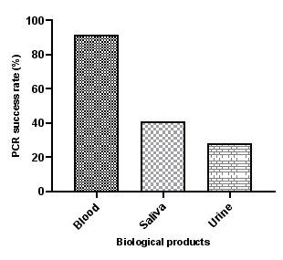
Figure 1. PCR success rate as a function of the biological product.
Saliva, urine and blood DNA extracts from these patients were subjected to nested PCR in separate batches using primers specific to the pfcrt, pfdhfr and pfK13 propeller genes. The amplification products of these genes were then loaded into adjacent lanes for each patient during agarose gel electrophoresis to highlight the amplification products.
The success rate of Plasmodium falciparum genomic DNA amplification by PCR was 41.13% (116/282) and 28.01% (79/282) respectively in saliva and urine compared with 91.84% (259/282) in blood (Figure 1). A significant difference (p < 0.05) was observed between the levels of amplification product in saliva and blood extracts and also between those (levels of amplification in urine and blood extracts).
3.2. Gene Detection Rate by Biological Product
Saliva, urine and blood extracts were subjected to nested PCR in separate batches using primers specific to the pfcrt, dhfr and K13 propeller genes. Agarose gel electrophoresis of the amplification products of each gene fragment detected Pfcrt at 80.85% (76/94), Pfdhfr at 95.74% (90/94) and PfK13 Propeller at 98.93% (93/94) in blood. In saliva, gene detection levels were 50% (47/94), 69.14% (65/94) and 4.26% (4/94) respectively for the K13 propeller, Pfdhfr and Pfcrt genes. Unlike the Pfcrt gene, which was not detected, the PfK13 Propeller and pfdhfr genes were detected in 45.74% (43/94) and 38.30% (36/94) of the urine respectively (Figure 2).
With regard to the amplification of the PfK13 Propeller gene, there was no significant difference between the detection rates of this gene in relation to the biological product. However, a significant difference (p-value<0.05) was observed between the detection rates of the Pfdhfr gene in the three biological products (blood, saliva and urine).
Concerning the Pfcrt gene, a significant difference was observed between the detection rates of this gene in blood and those (detection rates) in saliva and urine (p-value<0.05). However, no significant difference was observed between the detection rates of the pfcrt gene in saliva and those (detection rates) in urine.
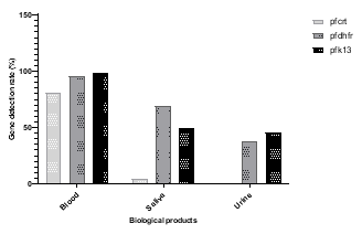
Figure 2. Detection rate of gene fragments by biological product.
3.3. Evaluation of Blood, Saliva and Urine as Biological Material for the Molecular Diagnosis of Malaria
We performed an analysis based on the presence or absence of Plasmodium falciparum DNA amplification products extracted from the blood, urine and saliva of patients with uncomplicated malaria. In relation to the population studied, the detection or non-detection of the pfcrt, pfdhfr and pfK13 propeller genes was assessed in each biological product. By associating the genes on the basis of their presence or absence in each biological product, the analysis revealed two trends suggesting that blood is the best biological sample for the molecular diagnosis of malaria using the pfcrt, pfdhfr and pfK13 propeller markers. However, the same analysis showed a close match between saliva and blood in the detection of the pfk13 propeller and pfdhfr genes in malaria patients (Figure 3). Guided by these observations, we performed an analysis of variance between blood, saliva and urine for the expression of the pfcrt, pfdhfr and pfK13 propeller genes in patients with uncomplicated malaria. This analysis identified two groups of biomarkers; (i) pfK13 propeller, pfdhfr and pfcrt and (ii) pfK13 propeller and pfdhfr demonstrated in blood and saliva respectively, while identifying them as satisfactory systems for discriminating malaria patients (p-value=0.03). Overall, the analysis suggested that unlike urine, blood and saliva would be the most appropriate biological samples for the molecular diagnosis of malaria (p-value <0.05).
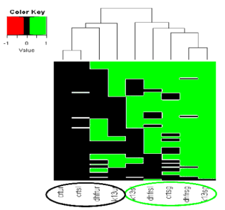
Figure 3. Graphical monitoring of the expression of the pfK13 propeller, pfdhfr and pfcrt genes in blood, saliva and urine extracts for the molecular diagnosis of malaria.
3.4. Comparative Analysis of the Performance of Saliva, Urine and Blood for the Detection of Antimalarial Drug Resistance Genes
Comparative analysis of the detectability of the pfK13 propeller, pfdhfr and pfcrt genes in blood, saliva and urine.
Principal component analysis was performed to assess the detectability relationship between the pfdhfr, pfK13 propeller and pfcrt genes in the blood, saliva and urine of subjects with uncomplicated malaria.
Figure 4A shows that there is an apparent correlation between: (i) the pfdhfr, pfK13 propeller and pfcrt genes in blood (PC1 axis (21.6%)); (ii) the pfdhfr and pfK13 propeller genes in urine (PC1 parameter (21.6%)) and in saliva (PC2 parameter (16.3%)); (iii) parasite density (PD) and the pfcrt gene in blood, the pfdhfr gene and the pfK13 propeller gene in saliva (parameters PC1 (21.6%) and PC2 (16.3%)).
In assessing the variability in the study population, analysis of Figure 4B shows the divergence between parasite density (PD) and urine and indicates that the pfk13 propeller and pfdhfr genes in urine may be responsible for the variability (PC1 and PC2 explaining more than 50% of the variability) in this study population (p < 0.05).
In addition, Pearson statistical tests (Table 2) showed a weak correlation between the pfdhfr and pfK13 propeller genes detected in blood and urine, in contrast to the pfcrt gene in blood (p = 0.09). Furthermore, these statistical tests excluded urine as a reliable biological product for the detection of the pfdhfr and pfK13 propeller genes in subjects with uncomplicated malaria (Table 2). However, the analysis confirmed that the detection performance of the pfdhfr and pfK13 propeller genes in saliva is close to that in blood. In summary, the analysis revealed that the detection of P. falciparum pfK13 propeller and pfdhfr genes in saliva could be a good alternative to blood for antimalarial drug resistance monitoring studies (Figure 4).
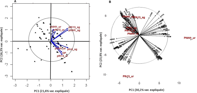
Figure 4. Comparative analysis of the relationship between the pfk13 propeller, pfdhfr, and pfcrt genes detected in blood, saliva and urine samples.
The acronyms ur, sl and sg stand for urine, saliva and blood respectively.
The PC1 and PC2 axes represent detectability and parasite density respectively.
Table 2. Pearson correlation between the pfdhfr, pfK13 propeller and pfcrt genes detected in blood, saliva and urine.

k13sg

k13sl

dhfrsg

dhfrsl

crtsg

DP

k13ur

dhfrur

k13sg

1

k13sl

0,10

1

dhfrsg

0,40*

-0,01

1

dhfrsl

-0,07

0,05

0,20*

1

crtsg

0,20*

0,30*

0,14

0,12

1

DP

0,05

0,17*

0,11

0,06

0,21*

1

k13ur

0,10

-0,04

0,07

0,01

0,12

-0,11

1

dhfrur

0,08

-0,01

0,11

-0,10

0,02

-0,07

-0,0004

1

Significant Pearson correlation value, p < 0.1
The figures in the table refer to the correlation coefficient.
Note: "ur", "sl" and "sg" refer to urine, saliva and blood respectively. "k13ur", "k13sl" and "k13sg" refer to the pfK13propeller gene in urine, saliva and blood respectively. The same applies to the pfdhfr and pfcrt genes in urine, saliva and blood.
Evaluation of saliva and urine performance by sensitivity, specificity, accuracy and positive predictive parameters.
The performance of saliva and urine samples was assessed for detecting pfdhfr, pfcrt and pfK13 molecular markers by ROC analysis assuming blood sample as reference . The data revealed a high sensitivity of saliva compared with urine in the detection of the pfdhfr, pfcrt and pfK13 genes (Table 3). Indeed, the pfdhfr gene in the saliva sample (pfdhfr_sl) recorded the highest sensitivity compared with the other genes. All the P. falciparum genes (pfdhfr, pfcrt and pfK13) detected in both urine and saliva (with the exception of the pfcrt gene in the urine sample) appeared to predict malaria infection well when blood biological sample is taken as the reference (positive predictive value > 0.80).
In addition, saliva sample showed better relative precision and specificity parameters assessing the detection performance of P. falciparum genes (Table 3). Furthermore, the ROC analysis confirmed and/or emphasised the detection of the pfK13 and pfdhfr genes in saliva as an efficient methodology that could substitute blood for antimalarial drug resistance surveillance studies. Finally, this analysis indexed the presence of the pfdhfr gene in saliva as the best parameter positively predicting malaria infection when blood is considered as the reference (Table 3).
Table 3. Evaluation of the performance of urine and saliva as biological samples for the study of genetic markers of chemo-resistance.

Parameters

Saliva

Urine

Pfk13

pfdhfr

pfcrt

Pfk13

pfdhfr

Pfcrt

Sensitivity

0,46

0,64

0,05

0,45

0,38

0

Specificity

0,2

0,5

0,5

0,5

0,5

0,01

Accuracy

0,43

0,54

0,05

0,45

0,38

0,01

Positive Predictive Value (PPV)

0,91

0,98

0,8

0,95

0,94

0

Comparative analysis of saliva and urine performances assessing malaria genetic biomarker fitting for malaria diagnostic.
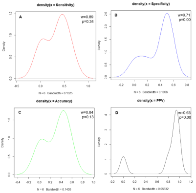
Figure 5. Shapiro normality test for assessing the distribution of urine and saliva samples statistical performance parameters.
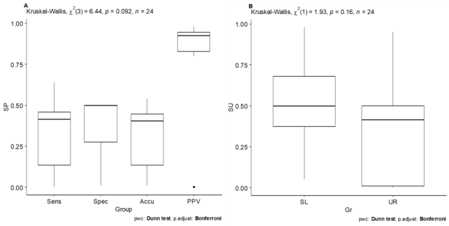
Figure 6. Kruskal-Wallis test evaluating statistical parameters variability (A) by comparing saliva and urine performances (B) in malaria molecular diagnosis procedure in alternative to blood sample.
Herein we compared statistical parameter performances as well as biological samples fitting well for malaria molecular diagnosis. Shapiro normality test exhibited normal distribution of sensibility and accuracy bio-statistical parameters weighing both saliva and urine biological samples performances, tested for substituting blood in malaria molecular diagnosis procedure (p>0.05) (Figure 5). Positive predictive values and specificity statistic parameters exhibited asymmetric distribution in analyzing saliva and urine performances. Interestingly, Kruskal-Wallis non-parametric statistical test suggested the positive predictive value parameter as a relative data variability source (p=0.09) (Figure 6A), making this parameter suitable for evaluating both saliva and urine biological samples performances differences. The same statistical test, by comparing saliva and urine samples statistical performance parameters, suggested a relative high performance of saliva sample as opposite to urine sample in detecting P. falciparum malaria biomarkers (p=0.16) (Figure 6B).
Sens, Spec, Accu and PPV acronyms referred to Sensitivity, Specificity, Accuracy and Positive Predictive Value statistical performances parameters. SL and UR referred saliva and urine samples respectively.
4. Discussion
Detection performance of plasmodial DNA in saliva, urine and blood
The malaria diagnostic tools developed to date all require blood to be taken. However, certain groups in the population are reluctant to take blood samples because of their cultural habits (blood taboo), or because of the fear linked to the trauma of the sting, especially when blood sampling is repeated. To overcome the obstacles associated with blood sampling, we need to think about palliative methods that can be used to meet all the needs of diagnosis and the study of cbemoresistance.
This study involved a comparative analysis of the amplification products of urine, saliva and blood DNA extracts with a view to finding an alternative to blood sampling in the molecular diagnosis of malaria and the study of genetic markers of antimalarial drug resistance. Analysis based on the presence or absence of Plasmodium falciparum DNA amplification products extracted from the blood, urine and saliva of subjects with uncomplicated malaria showed that the genomic DNA extracted from these biological products can be amplified to varying extents depending on the biological product. In fact, despite the low success rates of plasmodial DNA amplification products in urine (28.01%) and saliva (41.13%) compared with blood (91.84%), these two biological products (urine and saliva) appear to have real potential for studying antimalarial drug resistance genes. These rates although lower are close to those reported by Zahra et al, in Iran (95.8%, 47% and 29% respectively in blood, saliva and urine) where the level of transmission is similar. These results are consistent with those of Mharakurwa et al, in Zambia and Nwakanma et al, in Gambia who suggest that the sensitivity of molecular methods for malaria detection in saliva and urine is affected by several factors, namely DNA extraction methods, target gene size, sample fraction and sample preservation . These researchers reported that salivary DNA extraction with a commercial Qiagen kit had a 2.6-fold higher amplification success than the Chelex extraction that was used in this study. The low rates we obtained could therefore be explained by the method used in this study: the Chelex method for extracting plasmodial DNA.
The low amplification rate in saliva and urine may also be explained by the fact that the plasmodial DNA in these biological products is insufficient to provide a useful amplification model. This is because there is selective permeability for parasite DNA fragments. Mucosal membranes, such as the buccal mucosa and the epithelial lining of the oral cavity or bladder, act as filters that allow certain parasite constituents to pass outside the whole parasite . In addition, DNA from lysed parasites may passively enter saliva via serum or macrophage phagosomes via intra-oral bleeding or cervical gingival fluid . In addition, an ultrastructural pathological study of the kidney tissue of patients infected with P. falciparum revealed the presence of parasitized erythrocytes sequestered in glomerular and tubulointerstitial vessels and also immune complexes comprising IgG, C3 and malarial antigens . The release of plasmodial DNA into the urine could be a general phenomenon that occurs during infection. However, since Plasmodium DNA can be released into urine via a variety of possible routes, the actual route of entry has not yet been precisely defined . Although Plasmodium falciparum DNA has been successfully detected in saliva and urine samples, the precise mechanisms by whitch traces of parasites DNA appear in saliva and urine require further investigation.
However, whether or not the pfcrt, pfdhfr and pfK13 propeller genes were detected in each biological product showed that all three genes were detectable in blood and saliva, while only two of these genes (pfdhfr and pfK13 propeller) were detectable in urine. These results suggest that, unlike urine, blood and saliva are suitable biological samples for the detection of the pfdhfr and pfK13 propeller genes for antimalarial resistance surveillance studies. Previous studies using primers specific for antimalarial resistance genes have shown that the genes detected in saliva or urine samples were identical to those found in the peripheral blood of the same individual . This suggests that urine and saliva can be used as biological products for the molecular diagnosis of malaria.
The results of the multivariate statistical analysis showed that there was a correlation between the biological products and the detection of genes in saliva, urine and blood. This analysis highlighted the groups of genes (i) pfK13 propeller, pfdhfr and pfcrt and (ii) pfK13 propeller and pfdhfr detected in blood and saliva respectively as a satisfactory system for discriminating the study population. The various principal component analyses combined with the analysis of table 2 showed urine to be one of the main sources of variability in the study population (patients with uncomplicated malaria), unlike saliva and blood. These results also indicated that the pfK13 propeller and pfdhfr genes detected in urine were the two factors that explained more than 95% of the variability observed between malaria patients. The results obtained in this study also suggest that, unlike urine, saliva has gene amplification rates close to those of blood. In addition, biplot analysis combining the two principal components (PC1 and PC2) indicates that the level of detection of the pfK13 propeller and pfdhfr genes in saliva is similar to that of the pfcrt gene in blood. Biplot analysis also suggested that blood and saliva performed equally well in detecting antimalarial drug resistance genes. These results were confirmed by statistical analysis of the Pearson correlation, which at the same time showed a negative correlation between the components of urine and those of blood and saliva. All these results show that saliva could be the best alternative to blood for the detection of antimalarial drug resistance genes. These results are similar to those of Kwannan and Danwang who observed that the molecular detection of plasmodial DNA in malaria subjects in urine was less sensitive than in saliva . This is thought to be related to the small amount of DNA template in urine compared to saliva and blood. Quantitative real-time PCR showed that the average amount of plasmodial DNA in blood was 600 and 2500 times higher than that in saliva and urine respectively .
Parasite density (PD) was then integrated as a control tool in the detection of molecular markers of antimalarial drug resistance. The analysis showed a low sensitivity of the parasite density parameter in the molecular diagnosis of malaria based on gene detection. In addition, the average parasite density of the different study sites was above 500 parasites/µL of blood; the threshold below whitch the effect of parasite density is noticeable in the molecular diagnosis of malaria . These results suggest that detection of the pfKk13 propeller and pfdhfr genes in saliva is poorly correlated with parasite density (PD). However, PD shows the same trends in terms of Pearson correlation with respect to blood and saliva unlike urine. This other result confirms that saliva can be used as a good alternative to blood for the detection of molecular markers of P. falciparum resistance to antimalarial drugs. According to the results of the ROC (receiver operational characteristic) analysis, in contrast to urine, saliva had better precision, specificity and positive prediction parameters, confirming the high performance and/or tendency of saliva to replace blood in the molecular diagnosis of malaria (based on the detection of antimalarial drug resistance genes). When blood was taken as the reference, the three genes pfK13 propeller, pfdhfr and pfcrt detected in saliva appeared to predict malaria infection well (positive predictive value > 0.80). In addition, the ROC analysis confirmed the detection of the pfK13 propeller and pfdhfr genes in saliva as an effective molecular tool for discriminating malaria patients as an alternative to blood sampling. Finally, the analysis indexed the presence of pfdhfr in saliva as the best parameter positively predicting malaria infection in the study population when the blood sample was considered as reference.
In addition, to consolidate our analysis, we used the Kruskal-Wallis test to compare the performance of statistical parameters and better assess the suitability of biological samples for the molecular diagnosis of malaria. This test suggested that the positive predictive value is a source of relative variability in the data (p=0.09), making this parameter suitable for assessing differences in performance between samples. Used subsequently to compare the performance of saliva and urine, this test suggested a relatively high performance of saliva compared to urine in the detection of molecular biomarkers of P. falciparum malaria (p=0.16). Ultimately, the analysis indicated that saliva was an appropriate biological product for the molecular diagnosis of malaria. For this study, the analysis also indexed the detection of the pfdhfr gene in saliva as the best parameter positively predicting malaria infection in the population studied when the blood sample was taken as a reference.
5. Conclusion
Current malaria diagnostic tools all require blood sampling, which is still not accepted by patients. Given this situation, it was necessary to explore alternative non-invasive tools such as saliva and urine for malaria diagnosis. The aim of this study was to evaluate the performance of saliva and urine in detecting the molecular markers of Plasmodium falciparum resistance to antimalarial drugs (the pfdhfr, pfcrt and pfK13 propeller genes). Thus, taking blood as the reference biological product, our study showed that saliva is a high-performance biological product that can potentially be used as an alternative non-invasive sample for the study of molecular markers of Plasmodium falciparum resistance to antimalarial drugs. In fact, the levels of detection of molecular markers of resistance to antimalarial drugs studied in saliva are close to those in blood.
Abbreviations

Pfcrt

Plasmodium Falciparum Chloroquine Resistance Transporter

Pfdhfr

Plasmodium Falciparum Dihydrofolate Reductase

Pfk13 propeller

Plasmodium Falciparum Kelch13 Propeller

PCR

Polymerase Chain Reaction

DNA

Deoxyribonucleic Acid

ELISA

Enzyme-Linked Immuno-Sorbent Assay

EDTA

Ethylene Diamine Tetra-Acetic Acid

Ethical Considerations
The study was conducted in accordance with the Declaration of Helsinki and approval was received from the National Ethics and Research Committee (CNER) of the Côte d'Ivoire Ministry of Health and AIDS. Following appropriate information and explanations, written consent was obtained from the adult participants and the parents or legal guardians of all children wishing to participate in the study prior to sampling.
Acknowledgments
The authors express their deep gratitude to Professor Mireille DOSSO (Director of the Institut Pasteur de Côte d'Ivoire) who allowed them to use the equipment of the molecular biology platform of the Institut Pasteur de Côte d'Ivoire to perform the PCR tests. The authors would also like to thank the staff at the study sites (Anonkoua-kouté, Port-Bouët and Ayamé) for their efforts and cooperation in recruiting patients and collecting samples.
Conflicts of Interest
The authors declare no conflicts of interest.
References
[1] WHO. World Malaria Report 2023: Information pack, regional data and trends. [online]. Available:
[2] WHO, T3: test. treat. track. Scaling up diagnostic testing, treatment and surveillance for malaria. [online]. Available:
[3] Graz B, Willcox M, Szeless T, et al.. 'Test and treat' or presumptive treatment for malaria in high transmission situations? A reflection on the latest WHO guidelines. Malaria Journal, 2011; 10: 136.
[4] Putaporntip C., Buppan P., Jongwutiwes S., Improved performance with saliva and urine as alternative DNA sources for malaria diagnosis by mitochondrial DNA-based PCR assays. Clinical Microbiology and Infection, 2011, 17: 1484-1491.
[5] Boahen O, Owusu-Agyei S, Febir LG, et al. Community perception and beliefs about blood draw for clinical research in Ghana. Transactions of the Royal Society of Tropical Medicine and Hygiene, 2013; 107: 261-5.
[6] Chatio S, Baiden F, Achana FS, et al. Knowledge and perceptions about clinical trials and the use of biomedical samples: findings from a qualitative study in rural Northern Ghana. PLoS One, 2016; 11: e0152854.
[7] Baiden F, Owusu-Agyei S, Okyere E, et al. Acceptability of rapid diagnostic test-based management of malaria among caregivers of Under-Five children in rural Ghana. PLoS One, 2012; 7: e45556.
[8] Newton S, Doku V, Geissler W, et al. Drawing blood from young children: lessons learned from a trial in Ghana. Transactions of the Royal Society of Tropical Medicine and Hygiene, 2009; 103: 497-9.
[9] Achieng F, Rosen JG, Cherop RY, et al. Caregiver and community perceptions and experiences participating in an infant malaria prevention trial of PfSPZ vaccine administered by direct venous inoculation: a qualitative study in Siaya County, Western Kenya. Malaria Journal, 2020; 19: 226.
[10] Mharakurwa S., Simoloka C., Thuma P. E., Shiff C. J., Sullivan D. J., PCR detection of Plasmodium falciparum in human urine and saliva samples. Malaria Journal, 2006, 5: 103.
[11] Nwakanma D. C., Gomez-Escobar N., Walther M., Crozier S., Dubovsky F., Malkin E., Locke E., Conway D. J., Quantitative detection of Plasmodium falciparum DNA in saliva, blood, and urine. Journal of Infectious Diseases, 2009, 199(11): 1567-74.
[12] Aninagyei E, Abraham J, Atiiga P, et al. Evaluating the potential of using urine and saliva specimens for malaria diagnosis in suspected patients in Ghana. Malaria Journal, 2020; 19: 349.
[13] Al-Shehri H, Power BJ, Archer J, et al. Non-invasive surveillance of Plasmodium infection by real- time PCR analysis of ethanol preserved faeces from Ugandan school children with intestinal schistosomiasis. Malaria Journal, 2019; 18: 109.
[14] Adja A. M., N'goran E. K., Koudou B. G., Dia I., Kengne P., Fontenille D., Chandre F., Contribution of Anopheles funestus, An. gambiae and An. nili (Diptera: Culicidae) to the perennial malaria transmission in the southern and western forest areas of Côte d'Ivoire. 2011, Annals of Tropical Medicine and Parasitology, 105: 13-24.
[15] Miguel R. H.; Coura J. R., Samudio F., Suárez-mutis M. C., Evaluation of three different DNA extraction methods from blood samples collected in dried filter paper in Plasmodium subpatent infections from the Amazon region in Brazil. Revista do Instituto de Medicina Tropical de São Paulo, 2013, 55(3): 205-8.
[16] Kain K. C. & Lanar D. E., Determination of genetic variation within Plasmodium falciparum by using enzymatically amplified DNA from filter paper disks impregnated with whole blood. Journal of Clinical Microbiology, 1991, 29: 1171-1174.
[17] Plowe C. V. & Wellems T. E., Molecular approaches to the spreading problem of drug resistant malaria. Advances in Experimental Medicine and Biology, 1995, 390: 197-209.
[18] R Core Team, A language and environment for statistical computing. R Foundation for Statistical Computing, Vienna, Austria. ISBN 3-900051-07-0, URL accessed on 18 February 2017 at
[19] Noel D. D., Inza J. F., Nafan D., Mohamed L. B., Jean-Luc A. M., Olefongo D., Loukou N. E., Martial D. S. Y., Souleymane S., Giovanni M., A Quick Computational Statistical Pipeline Developed in R Programing Environment for Agronomic Metric Data Analysis. American Journal of Bioinformatics Research 2019, 9(1): 22-44
[20] Dago Dougba Noel, Lallié Hermann Désiré N. M., Diarrassouba Nafan, Silué Souleymane and Giovanni Malerba. Correlation between accuracy sensitivity specificity and positive predictive value parameters in detecting differentially expressed genes. International Journal of Development Research, 2015, Vol. 5, Issue, 03, pp. 3734-3739, March, 2015. Available online at
[21] Zahra G. N., Hormozd O., Lame A., Ahmad R. M., Ahmad R., Zahra R., Mehdi N., Elham R., Mitochondrial PCR-based malaria detection in saliva and urine of symptomatic patients. Transactions of the Royal Society of Tropical Medicine and Hygiene, 2014, 108: 358-362.
[22] Kwannan Nantavisai, Malaria detection using non-blood samples. Songklanakarin. Journal of Science and Technology, 2014, 634 36 (6): 633-641.
[23] A-Elgayoum, S. M., El-Rayah, E.-A., Giha, H. A., Towards a noninvasive approach to malaria diagnosis: detection of parasite DNA in body secretions and surface mucosa. Journal of Molecular Microbiology and Biotechnology, 2010, 18, 148-155.
[24] Sutherland C. J., Hallett, R., Detecting malaria parasites outside the blood. Journal of Infectious Diseases, 2009, 199, 1561-3.
[25] Nguansangiam S., Day N. P., Hien T. T., Mai N. T., Chaisri U., Riganti M., Dondorp A. M., Lee S. J., Phu N. H., Turner G. D., White N. J., Ferguson D. J., Pongponratn E., A quantitative ultrastructural study of renal pathology in fatal Plasmodium falciparum malaria. Tropical Medicine and International Health, 2007, 12, 1037-50.
[26] Das B. S., Renal failure in malaria. Journal of Vector Borne Diseases. 2008, 45, 83-97.
[27] Kawai Satoru, Megumi Sato, Naoko Kato-Hayashi, Hisashi Kishi, Michael A. Huffman, Yoshimasa Maeno, Richard Culleton, Shusuke Nakazawa, Detection of Plasmodium knowlesi DNA in the urine and faeces of a Japanese macaque (Macaca fuscata) over the course of an experimentally induced infection. Malaria Journal, 2014, 13: 373.
[28] Danwang C, Noubiap JJ, Souopgui J, et al. Accuracy of malaria diagnostic tests performed on noninvasively collected samples: a systematic review and metaanalysis. BMJ Global Health, 2021; 6: e005634.
[29] Bousema T., Okell L., Felger I., Drakeley C., Asymptomatic malaria infections: detectability, transmissibility and public health relevance. Nature Reviews Microbiology, 2014, 12(12): 833-840.
Cite This Article
  • APA Style

    Oléfongo, D., Noél, D. D., Angélo, K. K. B., Bérenger, A. A. A. A., Kouakou, B., et al. (2024). Assessment of Saliva and Urine Performance for Antimalarial Drug Resistance Molecular Markers Study. International Journal of Microbiology and Biotechnology, 9(3), 68-78. https://doi.org/10.11648/j.ijmb.20240903.14

    Copy | Download

    ACS Style

    Oléfongo, D.; Noél, D. D.; Angélo, K. K. B.; Bérenger, A. A. A. A.; Kouakou, B., et al. Assessment of Saliva and Urine Performance for Antimalarial Drug Resistance Molecular Markers Study. Int. J. Microbiol. Biotechnol. 2024, 9(3), 68-78. doi: 10.11648/j.ijmb.20240903.14

    Copy | Download

    AMA Style

    Oléfongo D, Noél DD, Angélo KKB, Bérenger AAAA, Kouakou B, et al. Assessment of Saliva and Urine Performance for Antimalarial Drug Resistance Molecular Markers Study. Int J Microbiol Biotechnol. 2024;9(3):68-78. doi: 10.11648/j.ijmb.20240903.14

    Copy | Download

  • @article{10.11648/j.ijmb.20240903.14,
      author = {Dagnogo Oléfongo and Dago Dougba Noél and Kouman Kouamé Bouatini Angélo and Ako Ako Ako Aristide Bérenger and Bla Kouakou and Touré Offianan André and Djaman Allico Joseph},
      title = {Assessment of Saliva and Urine Performance for Antimalarial Drug Resistance Molecular Markers Study
    },
      journal = {International Journal of Microbiology and Biotechnology},
      volume = {9},
      number = {3},
      pages = {68-78},
      doi = {10.11648/j.ijmb.20240903.14},
      url = {https://doi.org/10.11648/j.ijmb.20240903.14},
      eprint = {https://article.sciencepublishinggroup.com/pdf/10.11648.j.ijmb.20240903.14},
      abstract = {Background: The malaria diagnostic tools developed to date require blood to be taken. However, certain groups in the population are reluctant to take blood samples because of their cultural habits (blood taboo), or because of the fear associated with the trauma of the injection, especially when the sample is taken repeatedly. Saliva and urine, which are not very invasive to collect, have not been widely used for malaria diagnosis. The aim of this study is to assess the performance of saliva and urine in detecting molecular markers of Plasmodium falciparum resistance to antimalarial drugs. Methodology: Blood, urine and saliva samples were collected in three different localities from 94 patients over 2 years of age with microscopically confirmed Plasmodium falciparum uncomplicated malaria. P. falciparum genomic DNA (Deoxyribonucleic acid) was then extracted and amplified using primers specific for the Pfcrt (Plasmodium falciparum Chloroquine Resistance Transporter), Pfdhfr (Plasmodium falciparum dihydrofolate reductase) and PfK13 propeller (Plasmodium falciparum Kelch13 propeller) genes. The amplification products were processed by electrophoresis and analyzed against blood, saliva and urine samples. A multivariate statistical analysis in R programming environment was performed aiming to assess the performance of blood, saliva and urine samples in detecting molecular markers of P. falciparum resistance. Results: Agarose gel electrophoresis of the amplification products of each gene detected the Pfcrt genes at 80.85% (76/94), Pfdhfr at 95.74% (90/94) and PfK13 Propeller at 98.93% (93/94) in blood. In saliva, gene detection levels were 50% (47/94), 69.14% (65/94) and 4.26% (4/94) respectively for the K13 propeller, Pfdhfr and Pfcrt genes. Unlike the Pfcrt gene, which was not detected, 45.74% (43/94) and 38.30% (36/94) of PfK13 Propeller and pfdhfr genes respectively were detected in urine. Taking blood as the reference biological sample, statistical analysis showed that unlike urine, saliva exhibited a detection performance for molecular markers of antimalarial drug resistance (pfcrt, pfdhfr, pfK13 propeller) close to that of blood (p pfdhfr, pfcrt and pfK13 using ROC (receiver operational characteristic) analysis. The data revealed a high sensitivity of saliva compared with urine in the detection of the pfdhfr, pfcrt and pfK13 propeller genes. Conclusion: The levels of detection of molecular markers of antimalarial drug resistance studied in saliva are close to those in blood. Saliva is a high-performance biological product that could potentially be used as an alternative non-invasive sample for the study of molecular markers of Plasmodium falciparum resistance to antimalarial drugs.
    },
     year = {2024}
    }
    

    Copy | Download

  • TY  - JOUR
    T1  - Assessment of Saliva and Urine Performance for Antimalarial Drug Resistance Molecular Markers Study
    
    AU  - Dagnogo Oléfongo
    AU  - Dago Dougba Noél
    AU  - Kouman Kouamé Bouatini Angélo
    AU  - Ako Ako Ako Aristide Bérenger
    AU  - Bla Kouakou
    AU  - Touré Offianan André
    AU  - Djaman Allico Joseph
    Y1  - 2024/08/20
    PY  - 2024
    N1  - https://doi.org/10.11648/j.ijmb.20240903.14
    DO  - 10.11648/j.ijmb.20240903.14
    T2  - International Journal of Microbiology and Biotechnology
    JF  - International Journal of Microbiology and Biotechnology
    JO  - International Journal of Microbiology and Biotechnology
    SP  - 68
    EP  - 78
    PB  - Science Publishing Group
    SN  - 2578-9686
    UR  - https://doi.org/10.11648/j.ijmb.20240903.14
    AB  - Background: The malaria diagnostic tools developed to date require blood to be taken. However, certain groups in the population are reluctant to take blood samples because of their cultural habits (blood taboo), or because of the fear associated with the trauma of the injection, especially when the sample is taken repeatedly. Saliva and urine, which are not very invasive to collect, have not been widely used for malaria diagnosis. The aim of this study is to assess the performance of saliva and urine in detecting molecular markers of Plasmodium falciparum resistance to antimalarial drugs. Methodology: Blood, urine and saliva samples were collected in three different localities from 94 patients over 2 years of age with microscopically confirmed Plasmodium falciparum uncomplicated malaria. P. falciparum genomic DNA (Deoxyribonucleic acid) was then extracted and amplified using primers specific for the Pfcrt (Plasmodium falciparum Chloroquine Resistance Transporter), Pfdhfr (Plasmodium falciparum dihydrofolate reductase) and PfK13 propeller (Plasmodium falciparum Kelch13 propeller) genes. The amplification products were processed by electrophoresis and analyzed against blood, saliva and urine samples. A multivariate statistical analysis in R programming environment was performed aiming to assess the performance of blood, saliva and urine samples in detecting molecular markers of P. falciparum resistance. Results: Agarose gel electrophoresis of the amplification products of each gene detected the Pfcrt genes at 80.85% (76/94), Pfdhfr at 95.74% (90/94) and PfK13 Propeller at 98.93% (93/94) in blood. In saliva, gene detection levels were 50% (47/94), 69.14% (65/94) and 4.26% (4/94) respectively for the K13 propeller, Pfdhfr and Pfcrt genes. Unlike the Pfcrt gene, which was not detected, 45.74% (43/94) and 38.30% (36/94) of PfK13 Propeller and pfdhfr genes respectively were detected in urine. Taking blood as the reference biological sample, statistical analysis showed that unlike urine, saliva exhibited a detection performance for molecular markers of antimalarial drug resistance (pfcrt, pfdhfr, pfK13 propeller) close to that of blood (p pfdhfr, pfcrt and pfK13 using ROC (receiver operational characteristic) analysis. The data revealed a high sensitivity of saliva compared with urine in the detection of the pfdhfr, pfcrt and pfK13 propeller genes. Conclusion: The levels of detection of molecular markers of antimalarial drug resistance studied in saliva are close to those in blood. Saliva is a high-performance biological product that could potentially be used as an alternative non-invasive sample for the study of molecular markers of Plasmodium falciparum resistance to antimalarial drugs.
    
    VL  - 9
    IS  - 3
    ER  - 

    Copy | Download

Author Information
  • Abstract
  • Keywords
  • Document Sections

    1. 1. Introduction
    2. 2. Materials and Methods
    3. 3. Results
    4. 4. Discussion
    5. 5. Conclusion
    Show Full Outline
  • Abbreviations
  • Ethical Considerations
  • Acknowledgments
  • Conflicts of Interest
  • References
  • Cite This Article
  • Author Information